The American College of Radiology, the Society of Breast Imaging and other national medical organizations recommend that women 40 and older get yearly mammograms. The American Cancer Society agrees that getting yearly mammograms starting at age 40 has the most benefit; they urge women to start no later than age 45. Those at increased risk due to a family history or other factors should talk to their doctor about screening earlier. Women age 55 and older may choose to be screened every one to two years but, on average, yearly screening will find cancers earlier.
At Labette Health, we provide a full range of breast screening and diagnostic services such as:
Screening Mammogram
A screening mammogram is a two-dimensional picture of the breast and used to detect breast tumors in women who have no symptoms of breast cancer. It is still one of the most advanced tools available for detecting breast abnormalities. A screening mammo’s purpose is to detect tumors that are too deep/small to be detected any other way.
3D Mammography
Considered the most advanced technology in screening and diagnostic mammography, 3D mammography allows doctors to examine breast tissue by layer. Rather than viewing breast tissue in a flat image, as with 2D mammography, it reveals the fine details that previously could be hidden by the tissue above or below. 3D mammography allows for improved cancer detection—particularly in women with denser breasts.
3D Mammography has been proven to detect abnormalities 15 months earlier than a standard mammogram. Research also shows that 3D mammograms can increase early cancer detection rates and decrease call backs.
Diagnostic Mammogram
When a patient's screening mammogram requires further evaluation, a diagnostic mammogram is in order. The diagnostic evaluation serves as the next step to clarify any abnormalities detected during a screening mammogram or doctor's exam.
Breast Biopsy
Ultrasound-guided biopsy allows physicians to quickly and accurately get samples of breast tissue for evaluation after there has been a suspicious finding on a diagnostic mammogram. Ultrasound-guided biopsy enables surgeons to get a sample of breast tissue as small as your fingernail with high accuracy.
Breast Ultrasound
A radiation-free technique, breast ultrasounds generate images through sound waves. They can help distinguish normal breast tissue from a fluid-filled cyst or a solid mass that appears on a mammogram. Ultrasounds are typically used in conjunction with mammography; however, they do not replace mammography. It is not a screening method for breast cancer, but rather a diagnostic tool.
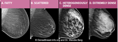
Dense Breasts
Breasts are made of fat and glands that make milk, held together by fibrous tissue. The more glands and fibrous tissue compared to fatty tissue that a woman has, the “denser” her breast tissue. Nearly half (over 40%) of women over age 40 have dense breasts.
Dense breasts have nothing to do with the way a breast looks or feels. Dense breast tissue increases the risk of developing breast cancer and of cancer being missed on the mammo.
When the radiologist looks at the mammo, they can see how dense the breasts are. In these four images, the dense tissue appears as white. Categories A and B are “not dense” breasts. Categories C and D are “dense” breasts.
Preparing for your mammogram
Here are some tips to help you prepare for your mammogram:
- Do not apply deodorant, perfume, talcum powder or lotion near the breast area, including underarms, on the day of your exam
- Let your physician or technologist know if there is a chance you may be pregnant
- Schedule your mammo after your menstrual cycle, when the breasts are less tender
- Wear a two-piece outfit on the day of your exam so it's easy to remove only your top for the exam